SAVED
File name .JPG
File alt. text
Image should be px wide x px tall.
Select Image
home - Biliary - Gallstone Disease - Biliary Colic Written by Dr Sebastian Zeki
Knowledge


Knows the physiology and biochemistry of bile and the pathogenesis
of gallstones

Is familiar with the normal anatomy and the anatomical variations of
the biliary tree

Recognises the symptoms and signs of the potential complications of
galllstone disease including biliary colic acute cholecystitis jaundice
due to calculous bile duct obstruction cholangitis and carcinoma

Knows the various techniques of diagnostic imaging including
ultrasound CT MRI ERCP EUS radionuclide techniques

Knows the various treatment options the indications for operative and
non-operative management and the risks of each

Knows the current national guidelines for use of ERCP and the risks
of the technique

Knows the ways in which gallbladder polyps are diagnosed and
managed

Knows that gallbladder and sphincter of Oddi dysfunction (SOD) may
account for otherwise unexplained abdominal pain

Recognises different types of SOD how they may present and how
they are investigated

Skills
Can select the most appropriate diagnostic and therapeutic
techniques for each clinical situation

Recognises possibility of diagnostic uncertainty in biliary dysmotility
and shows thoughtful judgement in each individual situation

Behaviours
Makes appropriate assessment stratifies urgency and plans
management of patients who have complications of gallstones

Biliary Colic

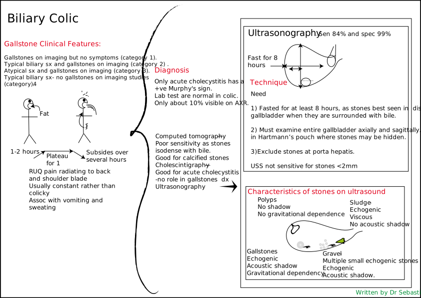
DiagnosisOnly acute cholecystitis has a +ve Murphy's sign.Lab test are normal in colic.Only about 10% visible on AXR. Gallstone Clinical Features:Gallstones on imaging but no symptoms (category 1).Typical biliary sx and gallstones on imaging (category 2) .Atypical sx and gallstones on imaging (category 3).Typical biliary sx- no gallstones on imaging studies (category 4). Need1) Fasted for at least 8 hours, as stones best seen in distended gallbladder when they are surrounded with bile.2) Must examine entire gallbladder axially and sagittally.Look in Hartmann’s pouch where stones may be hidden.3)Exclude stones at porta hepatis.USS not sensitive for stones <2mm Computed tomography — Poor sensitivity as stones isodense with bile.Good for calcified stonesCholescintigraphy — Good for acute cholecystitis -no role in gallstones dxUltrasonography Fast for 8 hours Poor if <1-2mm Biliary Colic Fat 1-2 hours RUQ pain radiating to back and shoulder bladeUsually constant rather than colickyAssoc with vomiting and sweating Plateau for 1 Subsides over several hours Characteristics of stones on ultrasound Gallstones EchogenicAcoustic shadow Gravitational dependency Gravel- Multiple small echogenic stones Echogenic Acoustic shadow. Sludge -EchogenicViscous No acoustic shadow Ultrasonography — Sen 84% and spec 99% PolypsNo shadowNo gravitational dependence Technique Written by Dr Sebastian Zeki

Related Stories

•Short-term prophylactic biliary stenting after complete common bile duct stone removal to prevent post-ERCP cholangitis in Japan (STEP-ERCP study): study protocol for a confirmatory multicentre randomised controlled trial

•Predicting gallstones risk with body composition analysis and machine learning: a dual-center cohort study

•Markedly Elevated CA 19-9 in Type I Mirizzi Syndrome in the Absence of Malignancy: A Case Report

•The biliary microbiome in symptomatic cholelithiasis: a prospective comparison across distinct bacterial niches

•Systematic review of gallbladder disorders: Molecular pathogenesis, biomarkers, diagnostic approaches, and emerging therapeutic strategies